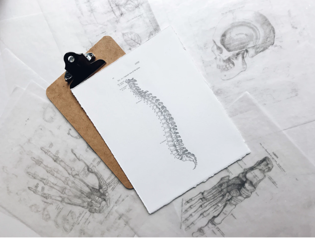
Spinal Fusion Surgery
Lumbar & Multilevel Spinal Fusion Surgery
A spinal fusion surgery is performed to stop motion at a painful vertebral segment. The purpose of this surgery is to reduce and allevaite discomfort generated from painful joints and degenerated discs.
TYPES OF SPINAL FUSION SURGERY
There are many different ways to perform a lumbar fusion, including anterior (through the abdomen), posterior (from behind) and lateral (through the side).
TECHNIQUES IN SPINAL FUSION SURGERY
Each spinal fusion surgery technique is designed to add bone graft either from the patient (auto graft) or from synthetic bone fillers. These grafts are added to the area to create a biological response similar to a fractured bone healing. The goal is to eliminate motion at the involved level and thus reduce pain in the process.
The main contributors to lumbar disc disease include:
- Degenerative disc disease
- Spondylolisthesis (meaning one vertebral body is slipping forward on the next vertebral body)
- Infection
- Tumor
- Scoliosis (curved spine)
How Spinal Fusion Surgery Works
Each level of the spine consists of the disc in front and two facets (joints) in the back. These structures work together to define a motion segment. When performing a fusion, for example L4 to L5, this is considered a one level fusion.
Nerve Relief
Commonly, most spinal fusions involve fusion of one or two levels. Motion tends to preserved in patients who will not notice much loss of motion in bending. Fusion rates are generally greater by 90% for nonsmokers and healthy individuals; risk for poor outcomem however, increases with smokers, diabetics, obesity, and immune-comprised individuals (rheumatoid arthritis, steroid usage, renal failure).
Multi-level Fusions
More than 2 level spinal fusions are indicated in patients with:
- Scoliosis
- Failed Back Surgery Syndrome
- Infection
- Trauma with associated spinal fractures
- Adjacent level deterioration
Types of Fusions
Posterior Lumbar Interbody Fusion (PLIF/TLIF) – Spinal fusion surgery is performed through the back and involves screws to stabilize the spine followed by cages (devices filled with bone) to stabilize the front of the spine.
Anterior Lumbar Interbody Fusion (ALIF) – done from the front and involves removing the entire disc and inserting cages filled with bone into the space.
Anterior and Posterior Fusion- fusion is done from the front and back.
Patients should be aware that with any spinal fusion surgery there is risk of clinical failure (meaning symptoms persist) despite achieving a solid fusion.
A successful result from spine fusion requires a number of factors:
- Accurate pre-operative work-up and diagnosis.
- Well trained spine surgeon.
- A patient who is willing to participate in a healthy life-style
- Non-smokers
- Ideal body weight
- Well controlled medical co-morbidities (diabetes, heart disease, and mental status)
The goal of any surgery is to have the surgeon and patient work closely together to get the best outcome. Well informed patients who are motivated in getting better will likely have better outcomes.
Anterior Cervical Discectomy and Fusion (ACDF)
Anterior Cervical Discectomy and Fusion (ACDF) is a surgical procedure to treat nerve root or spinal cord compression by decompressing the spinal cord and nerve roots of the cervical spine in order to stabilize the corresponding vertebrae. This procedure is used when other non-surgical treatments have failed. 90% of neck fusions are one and two level.
Anterior Cervical Discectomy
The nucleus pulposus (the jelly-like center of the disc) of the herniated disc bulges out through the annulus (surrounding wall) and presses on the nerve root next to it. This nerve root becomes inflamed and causes serious pain. The problem can also be caused by degenerative disc disease (spondylosis). The disc consists of about 80% water. When one grows older, the disc starts to dry out and shrink, causing small tears in the annulus and inflammation of the nerve root.
The spine surgeon enters the space between two discs through a small incision in front (= anterior) of and at the right or left side of the neck. The disc is completely removed, as well as arthritic bone spurs. The disc material, pressing on the spinal nerve or spinal cord, is then completely removed. The intervertebral foramen, the bone channel through which the spinal nerve runs, is then enlarged with a drill giving the nerve more room to exit the spinal canal.
To prevent the vertebrae from collapsing and to increase stability, the open space is often filled with bone graft, taken from the pelvis or cadaveric bone. The slow process of the bone graft joining the vertebrae together is called “fusion”. Sometimes a titanium plate is screwed on the vertebrae or screws are used between the vertebrae to increase stability during fusion, especially when there is more than one disc involved.
The surgery requires a short stay in the hospital (23 hour) and a gradual recovery between 1 to 2 weeks. The patient may be advised to wear a neck brace or collar (for up to 6 weeks) that serves to ensure proper spinal alignment. Wearing the brace heightens one’s awareness of posture and positioning and helps prevent movements (e.g., sudden and/or excessive bending or twisting of the neck) that may aggravate or slow down the healing process. It is especially advisable to wear a protective neck brace when traveling (e.g., by car), sleeping, showering, or any other activities in which the patient may not be able to ensure proper spinal alignment. In addition, physical therapy and related healing modalities (e.g., massage, acupuncture) may be recommended in order to promote proper healing, as well as to strengthen the surrounding muscles that can take over the neck brace’s ‘job’ of ensuring proper spinal alignment when the patient starts (around 4 to 6 weeks after surgery) to wean off the neck brace.
Spinal Fusion Surgery is a procedures that is used to correct issues with the vertebrea (the small bones in the spine). The procedures entails fusing together two or more vertebrae so they heal into a single, strong bone.